
What is Ultrasonography in Obstetrics?
Ultrasonography in obstetrics is a safe imaging test used during pregnancy. It uses sound waves to create pictures of the baby and the mother’s womb. Often, people call it a prenatal ultrasound or pregnancy scan. Doctors use this tool to check the baby’s growth and health. Because it is painless and does not use radiation, it is a common part of pregnancy care.
Why is Ultrasonography Important During Pregnancy?
Ultrasonography plays a key role in safe pregnancy monitoring. For example, it helps doctors spot problems early. It also lets parents see their baby’s first images. Most importantly, it gives doctors important details about the baby’s health. As a result, both mother and baby get the best care possible.
Common Uses and Benefits of Obstetric Ultrasound
Obstetric ultrasound offers many benefits for expecting parents. Here are some common uses:
Because of these benefits, doctors recommend regular ultrasounds during pregnancy. According to the World Health Organization (WHO), ultrasound helps improve pregnancy outcomes.
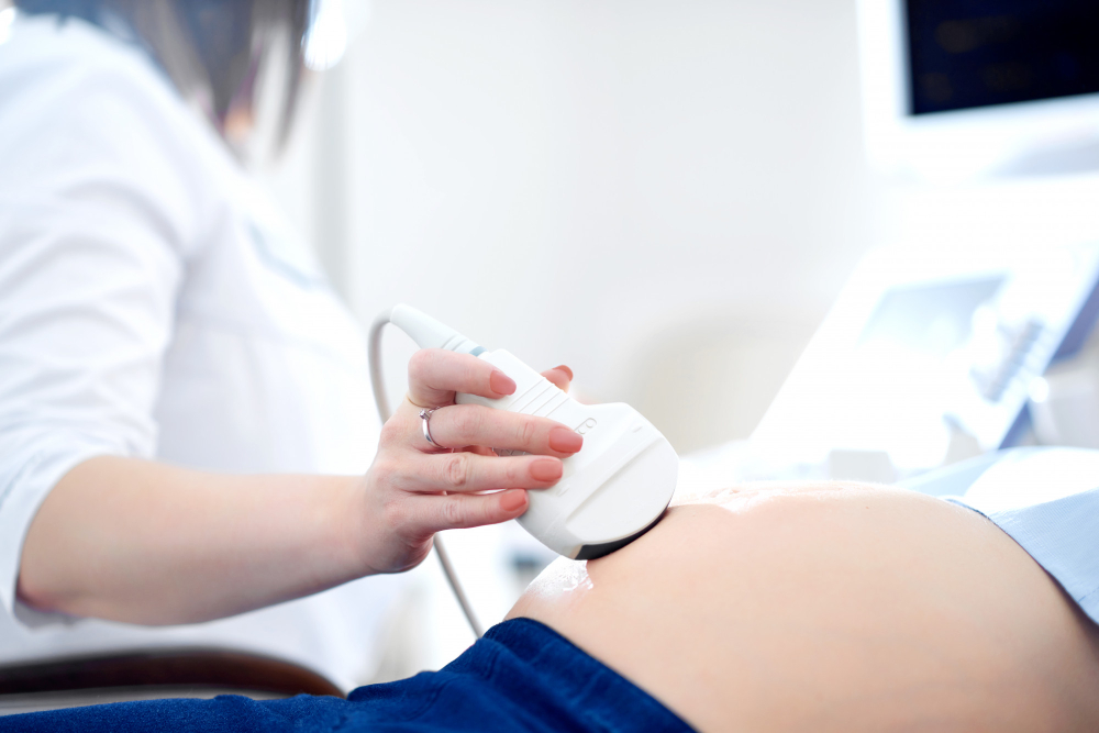
How Ultrasonography Works: The Procedure Explained
During an ultrasound, a trained technician or doctor will help you. First, you may lie down on a table. Next, a special gel is put on your belly. Then, a small device called a transducer moves over the skin. The device sends sound waves into the body. These waves bounce back and create images on a screen. The whole process is painless and usually takes less than 30 minutes. After the scan, you can return to normal activities right away.
Safety and Accuracy of Obstetric Ultrasound
Many parents worry about safety. However, prenatal ultrasound is very safe. It does not use harmful radiation. The Centers for Disease Control and Prevention (CDC) and WHO both say that ultrasound is safe for both mother and baby. In addition, ultrasound is accurate for checking the baby’s health, growth, and position. Still, it is important to have scans done by trained professionals.
When and How Often Should Pregnant Women Get Ultrasounds?
Most women have at least two ultrasounds during pregnancy. The first scan is often done in the first trimester, around 6–9 weeks. This scan confirms the pregnancy and checks the baby’s heartbeat. The second scan, called the anatomy scan, happens around 18–22 weeks. It checks the baby’s organs and growth. Sometimes, doctors may suggest more scans if needed. For example, extra scans may be needed if there are health concerns or if you are expecting twins. Always follow your doctor’s advice on the timing and number of scans.
Frequently Asked Questions About Pregnancy Ultrasound
Tips for Expecting Parents: Preparing for Your Ultrasound
Getting ready for your pregnancy scan is easy. Here are some helpful tips:
Remember, your doctor is there to help. If you feel nervous, talk to your healthcare team. They can explain each step and answer your questions.
Conclusion: The Value of Ultrasonography in Obstetrics
In summary, ultrasonography in obstetrics is a vital tool for safe pregnancy care. It helps doctors monitor your baby’s health and gives parents peace of mind. Because it is safe and accurate, it is a key part of modern prenatal care. For the best advice on pregnancy scans, consult your healthcare provider. They can guide you on when and how often to have ultrasounds for a healthy pregnancy.